 |
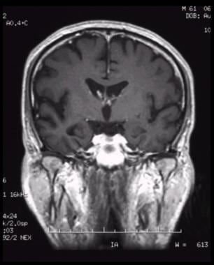
| Coronal, T1-weighted magnetic resonance imaging (MRI) scan in a patient with moderate Alzheimer disease. Brain image reveals hippocampal atrophy, especially on the right side. |
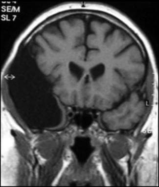 |
| Coronal T1-weighted MRI image through a brain lesion, showing homogeneity of the lesion, lack of a perceptible wall, lack of internal complexity, and CSF signal intensity. There is associated remodeling of the adjacent calvarium and brain displacement. These imaging features are typical of an arachnoid cyst. |
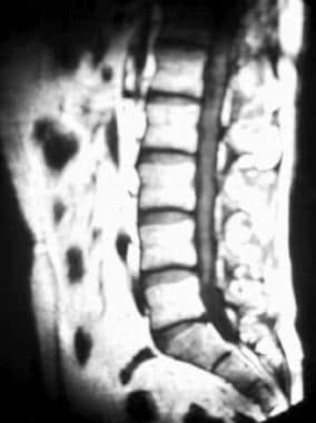 |
| Sagittal T1-weighted MRI of the lumbar spine in a patient with adhesive arachnoiditis who received epidural steroid injections. Image shows thickened and clumped nerve roots, which give the appearance of a tethered spinal cord. |
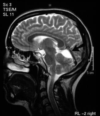 |
| T2-weighted sagittal MRI image of the brain in a 28-year-old woman with an incidental finding of a superior cerebellar cistern arachnoid cyst (arrow). |
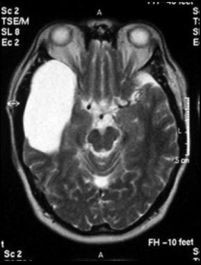 |
| Axial T2-weighted MRI image through the midbrain, showing a right middle cranial fossa homogeneous lesion with CSF signal intensity and no perceptible wall or internal complexity. There is associated remodeling of the adjacent sphenoid bone and brain displacement. These imaging features are typical of an arachnoid cyst. |
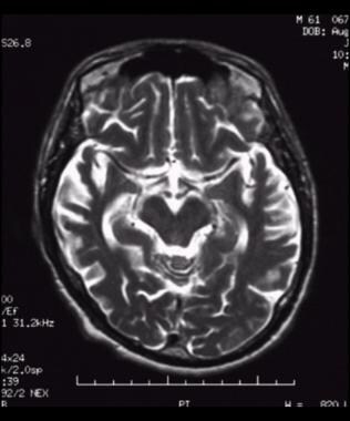 |
Axial, T2-weighted magnetic resonance imaging (MRI) scan shows bilateral temporal cortical atrophy with accentuated cortical sulci and dilated sylvian fissure; there is decreased involvement in the posterior aspect.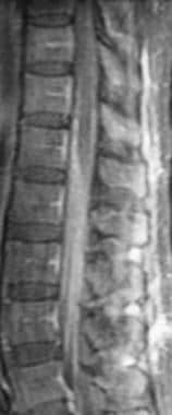 T1-weighted sagittal fat-suppressed contrast-enhanced MRI of the lumbar spine in tuberculous arachnoiditis and meningitis shows thin, linear leptomeningeal enhancement of the conus medullaris and cauda equina. 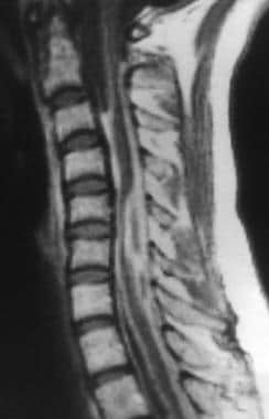 T1-weighted sagittal MRI of the cervical spine in tuberculous arachnoiditis shows nodular pockets of enhancement in the subarachnoid space after the administration of contrast material. 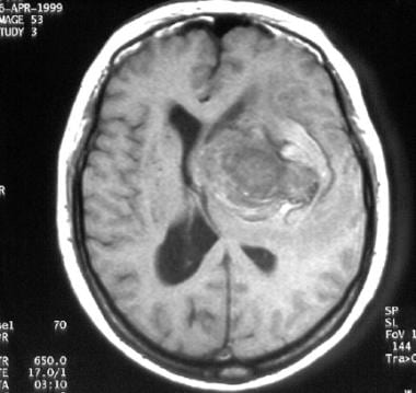 T1-weighted magnetic resonance image (MRI) of a middle-aged woman with progressive headaches, aphasia, and right-sided hemiparesis. A large intracerebral mass with a significant amount of surrounding edema is depicted. The lesion is a giant internal carotid artery aneurysm. 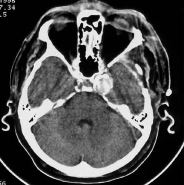 Nonenhanced CT scan of a middle-aged man with headaches. The patient had a giant aneurysm of the left internal carotid artery in its intracavernous segment. This aneurysm is densely calcified and is easily depicted. 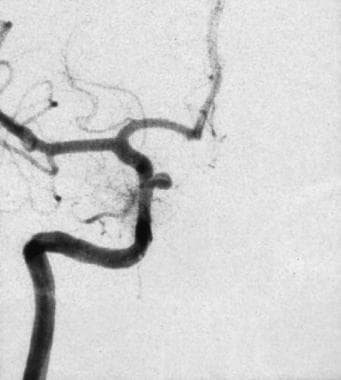 Left oblique cerebral angiogram in a patient with a proximal intracranial internal carotid artery aneurysm. The surgical approach to this aneurysm requires a craniotomy with an orbitotomy and drilling of the anterior clinoid process; however, this aneurysm has a favorable neck-to-fundus ratio for endovascular coil placement. 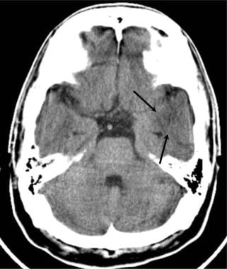 Brain abscess. Axial nonenhanced cranial CT scan in a patient who presented with fever, headache, and a previous paranasal sinus infection demonstrates a poorly defined pattern of mass effect and low attenuation in the left temporal lobe. The pattern is consistent with possible early cerebritis; however, glioma and infarct may have similar presentations. 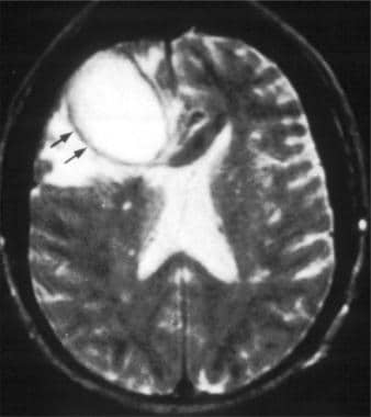 |
Brain abscess. Axial T2-weighted MRI in a patient with a right frontal abscess. Note the mass effect and surrounding edema. The wall of the abscess is relatively thin (black arrows).
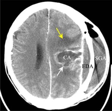
Brain abscess. Axial contrast-enhanced CT scan in a patient who was treated surgically for a depressed skull fracture. The left parietal cranial injury has become complicated by an abscess of the subgaleal space (SGA), of the epidural space (EDA), and within the left cerebral hemisphere (CA). Edema related to the abscess is indicated by the yellow arrow. The cerebral abscess wall enhances (white arrow).
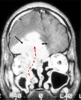
Brain abscess. Gadolinium-enhanced coronal T1-weighted MRI in a patient who presented with headache, fever, and diplopia. The right frontal lobe of the brain is shifted across the midline (double arrow) by an intracranial abscess (single black arrow) that has extended upward from the medial right orbit and medial ethmoid air cells (curved dotted arrow). Aspergillus organisms were recovered from the sinuses and brain tissue.